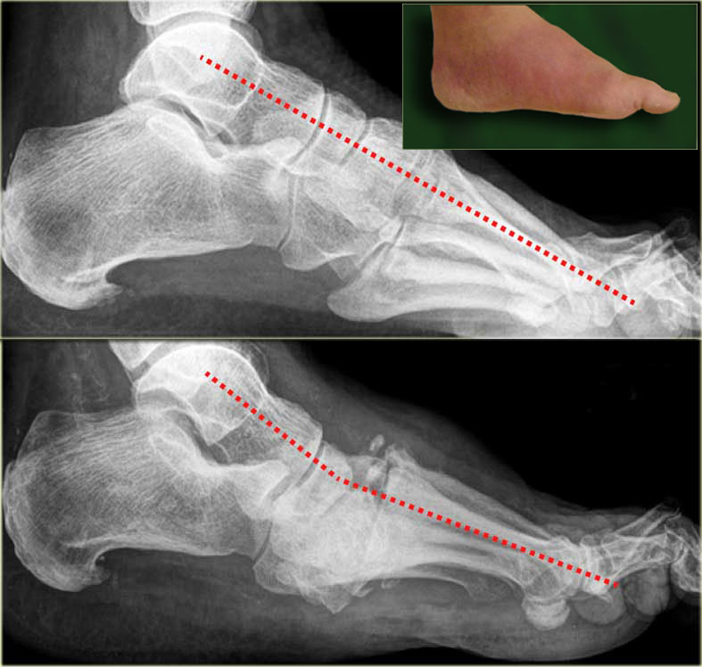
Periostal reaction and new bone formation at the 1st and 2nd metatarsals. Mnemonic - 6 Ds of charcot joint.

What The Radiologist Needs To Know About Charcot Foot Mautone 2015 Journal Of Medical Imaging And Radiation Oncology Wiley Online Library
It must be distinguished from other conditions such as osteomyelitis with efficiency and accuracy.

Charcot foot radiology findings. Modalities to differentiate between Charcots and infected foot is a challenge. However this classification does not cover the whole spectrum of CN. HIPAA compliance was observed.
The prognosis and treatment depends on it. MRI can be useful for early diagnosis monitoring disease activity and assessing complications such as infection. If there is bone marrow edema osteomyelitis is very likely.
Charcot neuro-osteoarthropathy of the foot has both an acute active and a chronic inactive phase. Many times the patient has been evaluated for a deep venous thrombosis and undergone oral or parenteral antibiotic therapy for cellulitis before a clinician obtains a radiograph or MRI that suggests the presence of Charcot arthropathy or osteomyelitis. Affected joints were examined for marrow articular periarticular and soft-tissue findings.
Charcot osteoarthropathy is a devastating process that occurs in the diabetic foot. Correlation of imaging findings and clinical symptoms. Charcot arthropathy typically presents with a warm swollen foot and ankle that cannot be differentiated easily from infection.
Imaging findings OR Procedure details Clinical presentation of Charcot neuro-osteo arthropathy in the foot. Destruction of articular cartilage. Charcot neuropathic osteoarthropathy can be classified by radiographic findings or anatomical location.
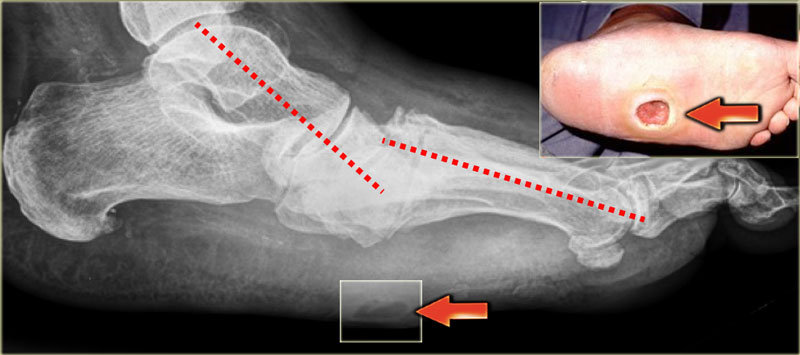
17 In 1966 Sydney Eichenholtz classified Charcot foot radiographically in three stages. No associated sinus tracks collections to suggest any secondary infection. Ulbrecht JS Wukich DK.
Dense bones subchondral sclerosis degeneration. Medical and surgical therapy. Radiographs may be normal or nondiagnostic during the early stage of Charcot foot.
Plain film radiographic findings can be normal in the. Currently the most widely recognised system is the Eichenholtz classification. Charcot joint involving foot is a progressive degenerativedestructive joint disorder in patients with abnormal pain sensation and proprioception.
Eur J Med Res. Destruction of the tarsometatarsal Lisfranc joint with dorsal subluxation of the medial cuneiform 1st and second metatarsals. Presence of superimposed osteomyelitis was documented.
Contrast-enhanced MR images in patients with diabetic neuropathic arthropathy of the foot were examined by two reviewers in consensus. Subchondral generalized osseous destruction involving mid foot with reduced tarsometatarsal joint space tarsal marrow edema and exuberant osteophytosis. Schlossbauer T Mioc T Sommerey S Kessler SB Reiser MF Pfeifer KJ.
Mnemonic - 6 Ds of Charcot joint. Dense bones subchondral sclerosis degeneration destruction of articular cartilage. Charcot with superimposed osteomyelitis.
To determine whether osteomyelitis in a Charcot foot at MR imaging is present follow the path of an ulcer or sinus tract to the bone and evaluate the signal intensity of the bone marrow. Sagittal T2 fat sat. A progressive degenerative destructive joint disorder in patients with abnormal pain sensation and proprioception.
Charcot progresses along four radiographically identifiable stages. Tech Foot Ankle Surg. It is most commonly secondary to diabetes.
Cellulitis and osteomyelitis commonly cause intense edema in the soft tissues and bone marrow of the foot. The acute Charcot foot is characterized by erythema edema and elevated temperature of the foot that can clinically mimic cellulitis or gout. Charcot joint involving foot.
Magnetic resonance imaging in early stage charcot arthropathy.

Licked Candy Stick Appearance Dd Leprosy Arthritic Psoriasis Neuropathic Joint Ballet Shoes Candy Sticks Sport Shoes

Diabetic Charcot Foot Orthoinfo Diabetes Foot Surgery Foot Deformities

Pdf What The Radiologist Needs To Know About Charcot Foot

Advanced Stage Of Neuro Osteo Arthropathy Charcot Foot On Download Scientific Diagram

Diabetic Charcot Foot Reconstruction Taylor Spatial Frame Knee Replacement Exercises Ankle Anatomy Knee Replacement

Domain Names On Twitter Radiology Student Radiology Medical Knowledge
Hip Radiographic Anatomy Wikiradiography Radiology Schools Anatomy Radiology Student

Pin On Radiography

Foot X Ray Normal Findings Bone And Spine Teeth Diseases Healthy Teeth X Ray

Charcot Joint Radiology Reference Article Radiopaedia Org

Charcot Arthropathy Radiology Case Radiopaedia Org

Conventional Radiology With Neuro Osteo Arthropathy Charcot Foot Of Download Scientific Diagram

Pin On Radiology

What The Radiologist Needs To Know About Charcot Foot Mautone 2015 Journal Of Medical Imaging And Radiation Oncology Wiley Online Library

Charcot Foot Radiology Case Radiopaedia Org
The Radiology Assistant Mri Examination

Charcot Foot Radiology Case Radiopaedia Org

What The Radiologist Needs To Know About Charcot Foot Mautone 2015 Journal Of Medical Imaging And Radiation Oncology Wiley Online Library
The Radiology Assistant Mri Examination
